Ultrasound-Guided Thoracentesis

Ultrasound-Guided Thoracentesis

Jailyn and Ben kick off a new podcast format where they discuss the full-length lectures published on the Core Ultrasound YouTube channel — adding commentary, clinical pearls, and behind-the-scenes discussion that doesn't make it into the edited videos.

Why this new podcast format? The YouTube videos are kept concise, but a lot of valuable clinical discussion gets left on the cutting room floor. This podcast fills that gap.

In this episode, they break down their approaches to ultrasound-guided thoracentesis, with a detour into related tips for paracentesis.

  • Static vs. Dynamic approach

    • Static: Identify the pocket, mark the spot, proceed with a landmark technique
    • Dynamic: Watch the needle in real-time as it enters the fluid
    • When to use each: Large effusions → static often sufficient; small or complex effusions → dynamic preferred
  • Identifying the right pocket

    • Find the largest fluid pocket that is cephalad enough to clear the diaphragm and caudal enough to avoid pneumothorax
    • Watch diaphragm excursion with respiration to confirm safe needle trajectory
    • Avoid inadvertent liver/spleen biopsy
  • Probe and needle technique for thoracentesis

    • Use a phased array or curvilinear probe to find the effusion, then switch to linear for the procedure
    • Orient transducer in the intercostal space (sagittal, rotated)
    • In-plane (long axis) approach recommended — short axis is mechanically difficult due to the ribs
    • Probe orientation: oblique when lateral, nearly horizontal when posterior
  • Paracentesis pearls

    • Out-of-plane technique can work well, given the larger pockets typical in the ED
    • Curvilinear probe makes needle identification harder vs. linear
    • For small-volume paras (e.g., ruling out SBP), dynamic approach is strongly preferred
  • Color Doppler before you needle

    • Scan the intended trajectory with color Doppler to rule out intercostal arteries or abdominal wall vessels (e.g., inferior epigastrics, caput medusa)
    • Release probe pressure nearly completely when looking for veins — they collapse easily and can be missed

Resources Mentioned

Thanks for listening — and happy scanning!

Denne episoden er hentet fra en åpen RSS-feed og er ikke publisert av Podme. Den kan derfor inneholde annonser.

Episoder(98)

The Other Side of the Heart - The Neglected Ventrcle

The Other Side of the Heart - The Neglected Ventrcle

In this episode, we discuss a video published on our YouTube channel on right heart ultrasound.    We discuss advanced methods to differentiate acute versus chronic right heart enlargement, with empha...

29 Jun 21min

Arthrocentesis in the Emergency Department

Arthrocentesis in the Emergency Department

In this episode, Ben and Jailyn discuss how they approach arthrotencesis in ED patients.  Watch the Youtube channel for a full run through for how to perform taps on the shoulder, knee, hip, elbow and...

16 Jun 25min

How to Save a Life with POCUS (with Dr. Sophia Fornbacher, MD)

How to Save a Life with POCUS (with Dr. Sophia Fornbacher, MD)

In this episode, we're thrilled to be joined by Dr. Sophia Fornbacher, ultrasound faculty and assistant professor at Riverside Community Hospital. Recently completing her ultrasound fellowship, Dr. Fo...

2 Jun 16min

The RUSH Exam - Undifferentiated Shock POCUS

The RUSH Exam - Undifferentiated Shock POCUS

When a patient rolls in hypotensive and tachycardic and you don't know why, the RUSH exam may be the most important tool in your hands. RUSH, Rapid Ultrasound in Shock and Hypotension, is a rapid, sys...

27 Mai 17min

Pericardial Effusion and When to Worry about Tamponade

Pericardial Effusion and When to Worry about Tamponade

Jailyn Avila and Ben Smith discuss a recent Core Ultrasound YouTube lecture on diagnosing pericardial effusions and cardiac tamponade with point-of-care ultrasound, emphasizing its importance in emerg...

19 Mai 16min
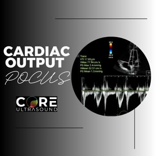
Measuring Cardiac Output Using Point-of-Care Ultrasound

Measuring Cardiac Output Using Point-of-Care Ultrasound

How to Obtain Cardiac Output (5 Minute Sono Series)   In this video, we'll guide you through the steps to calculate the cardiac output using the stroke volume and the patients heart rate. Learn how to...

26 Jan 6min
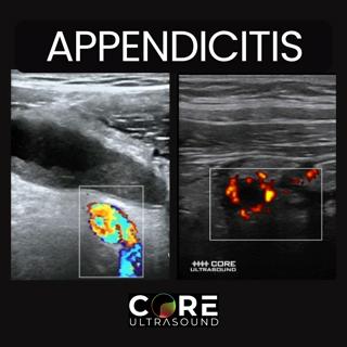
Appendicitis Diagnosis with Ultrasound

Appendicitis Diagnosis with Ultrasound

In this video, Jailyn explores the techniques and tips for diagnosing appendicitis using ultrasound. Learn about the preferred probes, patient preparation, and step-by-step guidance on locating the ap...

14 Jan 8min

Populært innen Fakta

fastlegen
dine-penger-pengeradet
relasjonspodden-med-dora-thorhallsdottir-kjersti-idem
foreldreradet
treningspodden
jakt-og-fiskepodden
mikkels-paskenotter
rss-strid-de-norske-borgerkrigene
rss-kunsten-a-leve
sinnsyn
hverdagspsyken
gravid-uke-for-uke
fryktlos
ryddepodden
rss-var-forste-kaffe
rss-sarbar-med-lotte-erik
tomprat-med-gunnar-tjomlid
babyverden
lederskap-nhhs-podkast-om-ledelse
rss-impressions-2